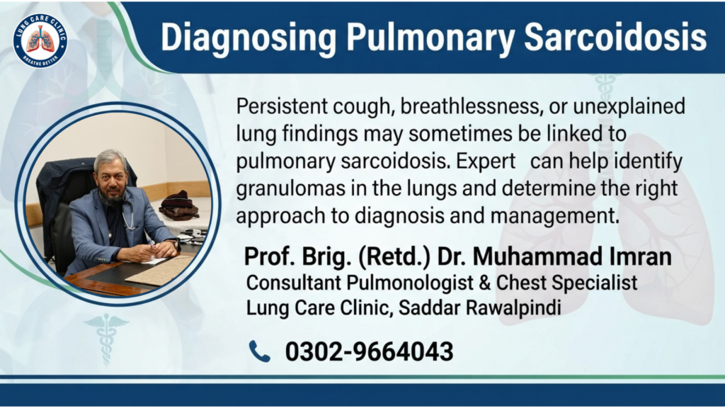
Sarcoidosis is an inflammatory condition that can affect different organs of the body, but the lungs and lymph nodes in the chest are among the most commonly involved areas. In pulmonary sarcoidosis, groups of inflammatory cells can form small collections called granulomas within lung tissue and nearby lymph nodes.
Some people with sarcoidosis have no noticeable symptoms, while others may develop persistent cough, shortness of breath, chest discomfort, fatigue, or reduced exercise tolerance. Because these symptoms can resemble other respiratory diseases, diagnosing pulmonary sarcoidosis requires careful evaluation rather than relying on a single symptom or test.
Understanding sarcoidosis lung disease, the role of granulomas in the lungs, and the diagnostic process can help patients recognize when specialist respiratory assessment may be necessary.
What Is Pulmonary Sarcoidosis?
Pulmonary sarcoidosis is sarcoidosis involving the lungs. It occurs when the immune system develops an abnormal inflammatory response that leads to the formation of granulomas.
A granuloma is a small organized collection of inflammatory cells. Granulomas are not unique to sarcoidosis. They can also occur with infections, certain immune conditions, and other diseases. Therefore, finding granulomas does not automatically confirm sarcoidosis.
This is one reason why diagnosing pulmonary sarcoidosis requires a combination of medical history, examination, imaging, laboratory testing, and sometimes tissue sampling.
What Causes Sarcoidosis?
The exact cause of sarcoidosis remains uncertain. Researchers believe that a combination of genetic and environmental factors may influence how the immune system responds to certain triggers.
Sarcoidosis is not considered a contagious disease. It can occur in people with no obvious history of lung disease.
Because its exact cause is not fully understood, doctors generally focus on identifying the pattern of organ involvement, excluding other possible causes, assessing disease severity, and determining whether treatment is necessary.
Granulomas in the Lungs
Granulomas are an important feature of sarcoidosis.
When inflammation occurs, immune cells can cluster together and form granulomas. In pulmonary sarcoidosis, these granulomas may develop within lung tissue or lymph nodes around the lungs.
The presence of granulomas can be identified through tissue examination, but their cause must still be established.
For example, tuberculosis and some fungal infections can also produce granulomas. This distinction is particularly important when evaluating patients in regions where tuberculosis is common.
Therefore, doctors may need to perform additional investigations before confirming sarcoidosis.
Common Symptoms of Sarcoidosis Lung Disease
Some patients with pulmonary sarcoidosis have minimal or no symptoms. Others develop respiratory symptoms that gradually affect daily activities.
Common symptoms may include:
- Persistent dry cough
- Shortness of breath
- Chest discomfort
- Wheezing in some patients
- Reduced exercise tolerance
- Fatigue
Symptoms can vary considerably from one person to another.
A persistent cough should not automatically be attributed to sarcoidosis because asthma, infections, allergies, reflux, COPD, and several other conditions can cause similar symptoms.
How Pulmonary Sarcoidosis Affects Breathing
Inflammation and granuloma formation can interfere with normal lung function.
In some patients, inflammation remains limited and lung function is preserved. In others, prolonged inflammation can lead to scarring or fibrosis, which may reduce the lungs’ ability to transfer oxygen efficiently.
As lung involvement becomes more significant, a person may notice increasing breathlessness during walking, climbing stairs, or physical activity.
This is why regular monitoring can be important for patients who have confirmed pulmonary sarcoidosis.
Diagnosing Pulmonary Sarcoidosis
There is no single test that confirms every case of sarcoidosis.
A pulmonologist usually begins with a detailed medical history and physical examination. The doctor may ask about respiratory symptoms, previous infections, occupational or environmental exposures, medications, and other health concerns.
Chest imaging is often an important part of the evaluation.
Chest X-Ray
A chest X-ray can identify enlarged lymph nodes, changes in lung tissue, or other abnormalities that may suggest sarcoidosis.
However, similar findings can occur in other diseases. Therefore, an abnormal X-ray does not by itself establish the diagnosis.
CT Scan of the Chest
A CT scan can provide more detailed information about the lungs and chest lymph nodes.
It may help doctors assess:
- Enlarged lymph nodes
- Small lung nodules
- Distribution of lung abnormalities
- Fibrotic or scar-like changes
- Other patterns of lung involvement
The imaging pattern can help guide further investigations.
Pulmonary Function Tests
Pulmonary function testing, including spirometry, can help determine how well the lungs are working.
Depending on the clinical situation, doctors may assess:
- Airflow
- Lung volumes
- Gas transfer
These measurements can help establish a baseline and monitor changes over time.
A patient may have significant abnormalities on imaging while having relatively preserved lung function, or symptoms may occur despite only modest imaging changes. This is why both clinical assessment and objective testing are valuable.
Blood Tests and Other Investigations
Blood tests may be used as part of the overall evaluation, although no single blood test can independently confirm pulmonary sarcoidosis.
Doctors may investigate markers of inflammation, calcium levels, organ function, and other findings based on the patient’s symptoms.
Additional testing may be recommended to look for involvement of organs outside the lungs.
Why Excluding Other Diseases Matters
One of the most important steps in diagnosing pulmonary sarcoidosis is ruling out conditions that can look similar.
Tuberculosis is particularly important to consider because it can cause granulomas, enlarged lymph nodes, cough, fever, weight loss, and other findings that may overlap with sarcoidosis.
Other infections, malignancies, autoimmune conditions, and occupational lung diseases may also need consideration.
A specialist therefore evaluates the complete clinical picture instead of diagnosing sarcoidosis based only on the presence of granulomas or an abnormal chest scan.
Role of Bronchoscopy and Tissue Sampling
In some patients, doctors may recommend bronchoscopy to investigate abnormalities inside the airways or obtain samples.
Bronchoscopy can sometimes allow the physician to collect fluid or tissue for laboratory and microscopic examination.
A biopsy may demonstrate the characteristic non-caseating granulomas associated with sarcoidosis. However, the results must be interpreted alongside clinical and imaging findings and after appropriate exclusion of infections and other causes.
Not every patient requires an invasive procedure. The decision depends on the individual clinical situation.
Does Every Patient Need Treatment?
Not necessarily.
Some patients with sarcoidosis have mild disease that remains stable or improves without specific treatment. Others may have progressive lung involvement or disease affecting important organs and may require treatment.
The decision depends on factors such as:
- Severity of symptoms
- Lung function
- Imaging findings
- Disease progression
- Involvement of other organs
- Risk of permanent organ damage
Treatment should therefore be individualized rather than based solely on the diagnosis.
Treatment of Pulmonary Sarcoidosis
When treatment is necessary, corticosteroids are commonly used to control significant inflammation. Other immune-modulating medicines may be considered when appropriate.
The treatment plan depends on the extent and severity of disease and the patient’s overall health.
Regular follow-up may include symptom assessment, pulmonary function testing, imaging, and monitoring for treatment-related effects.
When Should You See a Pulmonologist?
You should consider specialist respiratory evaluation if you have:
- Persistent unexplained cough
- Progressive breathlessness
- Unexplained abnormalities on a chest X-ray or CT scan
- Enlarged chest lymph nodes
- Known granulomas in the lungs
- Unexplained reduction in lung function
- Respiratory symptoms that persist despite initial treatment
Early specialist assessment can help distinguish sarcoidosis from other conditions with similar presentations.
Sarcoidosis lung disease is a complex inflammatory condition that can affect lung tissue and lymph nodes in the chest. Granulomas in the lungs are an important feature, but they can occur in several other diseases, making accurate diagnosis essential.
A comprehensive evaluation may include medical history, physical examination, chest imaging, pulmonary function testing, laboratory investigations, and, when appropriate, bronchoscopy or tissue sampling.
If you have persistent respiratory symptoms or unexplained lung abnormalities, do not assume the cause without proper evaluation. A pulmonologist can assess your symptoms, investigate possible causes, and develop an appropriate monitoring or treatment plan.
Prof. Brig. (Retd.) Dr. Muhammad Imran
Consultant Pulmonologist & Chest Specialist
Lung Care Clinic, Saddar Rawalpindi
0302-9664043