
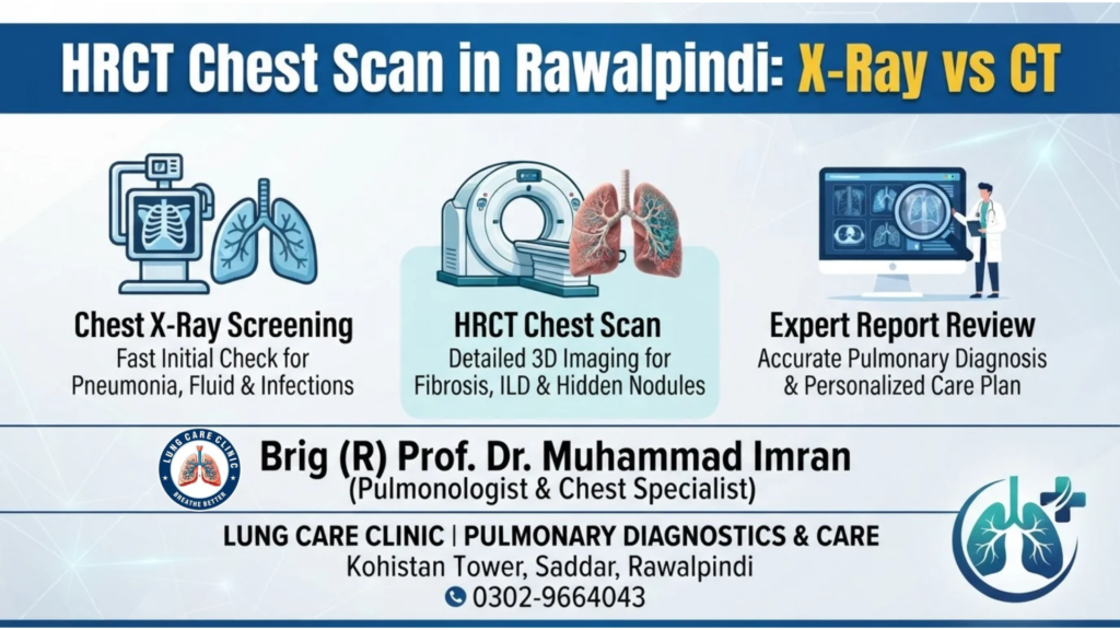
When evaluating persistent cough, unexplained breathlessness, or chest discomfort, medical imaging plays an essential role in identifying underlying lung conditions. Many patients wonder whether a standard chest radiograph provides enough diagnostic detail or if they require an HRCT chest scan in Rawalpindi for a deeper look inside their airways. Understanding how these two imaging tools work helps patients make informed decisions alongside their respiratory specialist.
Both tests use controlled X-ray beams to visualize the lungs, heart, ribs, and surrounding chest structures. However, their image clarity, level of anatomical detail, radiation exposure, and clinical applications differ significantly. Choosing the right diagnostic test at the right time prevents delayed diagnoses while avoiding unnecessary imaging.
Chest X-Ray vs HRCT Chest Scan: Key Differences
A standard chest X-ray and a High-Resolution Computed Tomography (HRCT) chest scan serve complementary roles in pulmonary medicine. While a chest X-ray offers a quick, broad overview of the chest cavity, an HRCT scan captures ultra-thin cross-sectional slices of lung tissue.
Image Detail and Resolution
A conventional chest X-ray compresses all three-dimensional structures of the chest—ribs, spine, heart, blood vessels, and lungs—onto a single flat two-dimensional image. Because organs overlap on a flat film, subtle inflammation or tiny lung nodules hidden behind the ribs or heart can remain invisible.
In contrast, an HRCT chest scan rotates around the body to capture hundreds of high-definition, millimeter-thin cross-sectional slices. A computer reconstructs these slices without overlapping shadows, allowing a pulmonologist to inspect tiny air sacs (alveoli), small bronchial airways, and delicate lung tissue walls with remarkable clarity.
Radiation Exposure
A single digital chest X-ray delivers a very low dose of radiation, comparable to a few days of natural background environmental exposure. An HRCT chest scan uses a higher radiation dose than a standard radiograph because it captures multiple detailed cross-sectional views. Modern low-dose HRCT protocols keep exposure strictly within safe diagnostic limits whenever high-resolution imaging is clinically justified.
Scan Duration and Procedure
A digital chest X-ray takes less than one minute while the patient stands against an imaging plate and holds their breath briefly. An HRCT chest scan takes roughly five to ten minutes while the patient lies comfortably on a motorized table that glides smoothly through a wide, open ring scanner. Standard HRCT chest scans rarely require intravenous contrast dye.
When Is a Standard Chest X-Ray Enough?
For most initial respiratory symptoms, a digital chest X-ray remains the ideal first-line diagnostic test. It is fast, widely accessible, cost-effective, and delivers immediate answers for many common acute chest problems.
A specialist typically starts with a chest X-ray to check for:
- Acute Pneumonia and Chest Infections: Identifies areas of fluid-filled lung consolidation caused by bacterial or viral pneumonia.
- Pleural Effusion: Detects moderate to large fluid accumulation in the space between the lungs and the chest wall.
- Pneumothorax (Collapsed Lung): Quickly reveals trapped air outside the lung requiring urgent care.
- Heart Size and Major Blood Vessels: Screens for cardiac enlargement or fluid overload contributing to shortness of breath.
- Routine Tuberculosis Screening and Treatment Follow-Up: Monitors visible upper-lobe lung shadows and tracks healing during medication courses.
When clinical symptoms match an straightforward infection and the chest X-ray clearly confirms the diagnosis, patients can begin targeted treatment immediately without needing a CT scan.
When Does a Pulmonologist Recommend an HRCT Chest Scan?
Sometimes a patient experiences persistent breathing symptoms, low oxygen levels, or abnormal lung function tests even though their standard chest X-ray appears completely normal. In these situations, ordering an HRCT chest scan in Rawalpindi provides the microscopic anatomical precision needed to uncover hidden lung conditions.
A chest specialist commonly recommends an HRCT chest scan in the following scenarios:
- Suspected Interstitial Lung Disease (ILD) and Pulmonary Fibrosis: Early lung tissue scarring appears as fine, lacy patterns that standard X-rays miss until scarring becomes advanced. HRCT detects ILD early so treatment can preserve lung function.
- Bronchiectasis Evaluation: Chronic productive cough with daily sputum often stems from permanently widened, scarred bronchial tubes. HRCT maps the exact extent and location of bronchiectasis across every lung lobe.
- Unexplained Chronic Cough or Persistent Breathlessness: When a cough lasts beyond eight weeks despite normal chest X-rays and initial therapy, HRCT rules out subtle airway disease, atypical infections, or early emphysema.
- Evaluating Lung Nodules or Shadows Seen on X-Ray: If a routine radiograph reveals a suspicious spot, an HRCT determines its exact size, borders, density, and location to guide safe monitoring or diagnostic bronchoscopy.
- Post-Infection or Post-Tuberculosis Lung Damage: Assesses residual airway narrowing, cavities, or localized scarring after severe pneumonia or tuberculosis treatment.
- Pre-Procedure Planning for Bronchoscopy: Provides a precise anatomical roadmap before performing bronchoalveolar lavage, bronchial biopsy, or transbronchial biopsy.
Comparing Both Diagnostic Tests Side by Side
| Feature / Scenario | Digital Chest X-Ray | HRCT Chest Scan |
|---|---|---|
| Primary Indication | Acute symptoms, routine screening, quick baseline checks | Chronic symptoms, unexplained abnormal tests, suspected early tissue disease |
| Dimensional Output | 2D single flat image (overlapping structures) | 3D multi-slice cross-sectional imaging |
| Tissue Resolution | Low to moderate; broad structural overview | Ultra-high; sub-millimeter anatomical detail |
| Radiation Dose | Very minimal | Higher, optimized via modern low-dose protocols |
| Need for Contrast Dye | None | Rarely needed for standard HRCT |
| Diagnostic Role | Best first-line screening tool for acute conditions | Best for fine tissue detail (fibrosis, bronchiectasis, small nodules) |
Understanding the practical strengths of each test clarifies why pulmonologists select one over the other based on your specific symptoms:
- Best First Step for Acute Symptoms: Standard Chest X-Ray quickly rules out pneumonia, large fluid collections, and collapsed lung.
- Best for Subtle Tissue & Airway Detail: HRCT Chest Scan detects early fibrosis, bronchiectasis, emphysema, and small nodules hidden on flat radiographs.
- Need for Contrast Dye: Neither a routine chest X-ray nor a standard HRCT chest scan typically requires contrast injection, making both safe for patients with kidney concerns.
- Clinical Value: Neither test replaces a thorough physical exam and spirometry; instead, imaging works alongside pulmonary function testing to form a complete diagnosis.
How to Prepare for a Chest X-Ray or HRCT Scan
Preparing properly ensures sharp, artifact-free images on your first visit:
- Remove Metallic Objects: Take off necklaces, chains, watches, and clothing with metal zippers, buttons, or snaps around the chest area, as metal creates streaks on images.
- Wear Loose, Comfortable Clothing: Cotton garments without metallic prints allow quick imaging without delays.
- Inform Staff About Pregnancy: Always notify the medical team before any X-ray or CT scan if there is any possibility of pregnancy.
- Bring All Previous Scans and Films: Carrying older chest X-rays or previous CT disks allows your pulmonologist to compare changes over time, often preventing unnecessary repeat scans.
Expert HRCT Chest Scan Evaluation in Rawalpindi
Imaging reports contain complex radiological terms such as ground-glass opacities, reticulonodular shadows, or traction bronchiectasis that can alarm patients when read without clinical context. An imaging scan is only as valuable as the specialist interpretation connecting those findings to your symptoms, oxygen levels, and lung function.
At Lung Care Clinic, located at Kohistan Tower, Saddar, Rawalpindi (Opposite AFIC), Brig (R) Prof. Dr. Muhammad Imran provides comprehensive pulmonary evaluation and direct review of both digital chest X-rays and HRCT chest scans.
By correlating high-resolution scan images with clinical history and spirometry testing, patients receive an accurate diagnosis, clear explanation of their lung health, and a targeted treatment plan without confusion or guesswork.
Frequently Asked Questions
Can a chest X-ray detect everything an HRCT chest scan shows?
No. A chest X-ray is a flat two-dimensional screening tool that can miss early lung scarring, mild emphysema, bronchiectasis, and tiny nodules. An HRCT chest scan captures thin cross-sectional slices that reveal fine lung tissue detail invisible on standard radiographs.
Does an HRCT chest scan require fasting or contrast injection?
A standard HRCT chest scan does not require contrast dye or fasting. Patients can eat, drink, and take their regular daily medications normally before the scan unless a specific contrast-enhanced CT is requested.
Should I get an HRCT chest scan before seeing a pulmonologist?
It is best to consult a pulmonologist first. A chest specialist will examine your lungs, review any existing X-rays, and determine whether an HRCT chest scan is truly necessary saving you unnecessary expense and radiation exposure.
For expert pulmonary consultation, chest imaging review, and personalized lung care, visit Lung Care Clinic at Kohistan Tower, Saddar, Rawalpindi (Opposite AFIC), or call 0302-9664043.